When you look at your teeth in a mirror, you can see a lot—but not everything. Some dental problems develop in places that are difficult to see, even when you have a clear view of your smile.
An intraoral camera gives dentists a closer, more detailed view of individual teeth and the surrounding areas. This small device can capture enlarged images inside the mouth, helping dental professionals identify concerns, document changes, and explain what they see to patients.
But what exactly can an intraoral camera show that a traditional mirror cannot?
A Much Closer View of Individual Teeth
A dental mirror provides a useful visual perspective, but an intraoral camera can capture highly magnified images of specific areas.
The camera is small enough to move around different parts of the mouth and can capture detailed views of individual teeth, gum tissue, fillings, crowns, and other structures.
These images can make subtle changes easier to observe and give the dental team a more detailed visual reference during an examination.
Areas That Are Difficult to See Directly
Some areas of the mouth can be challenging to inspect using a mirror alone. The camera can be positioned around the mouth to capture images from different angles.
This may allow the dentist to examine areas around the back teeth, along the gumline, or other surfaces that are difficult for patients to see themselves.
However, an intraoral camera does not replace other diagnostic tools. Problems between teeth or beneath the tooth surface may still require dental X-rays or other examinations.
Early Signs of Tooth Damage
An intraoral camera can help dentists document visible changes in the appearance of a tooth.
Small chips, cracks, worn areas, discoloration, or changes around an existing restoration may be easier to identify when the tooth is displayed as an enlarged image.
The camera does not diagnose every type of dental problem on its own, but it can provide valuable visual information that supports the overall examination.
Changes Along the Gumline
The gumline is another area where an intraoral camera can be useful.
Enlarged images may make visible signs such as redness, swelling, irritation, plaque accumulation, or other changes easier to observe.
Being able to see these areas on a screen can also help patients understand why their dentist may recommend improved brushing, flossing, professional cleaning, or additional evaluation.
The Condition of Existing Dental Work
An intraoral camera can also provide a closer look at existing dental restorations.
Fillings, crowns, bridges, and other restorations can be examined for visible wear, damage, changes around their edges, or other concerns.
Having detailed images can help the dentist compare the current appearance of a restoration with previous findings and determine whether closer monitoring or further evaluation may be appropriate.
A Better Way to Explain What Your Dentist Sees
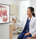
One of the biggest advantages of an intraoral camera is that it can make dental examinations more visual for patients.
Instead of simply being told that a dentist sees a worn area, irritated gum, or damaged restoration, patients may be able to see the area themselves on a monitor.
This can make conversations about oral health easier to understand. Patients can ask questions about specific areas and better understand why a particular treatment or preventive recommendation may be suggested.
Tracking Changes Over Time
Dental care is not only about identifying problems today. Monitoring changes over time is also important.
Intraoral images can provide a visual record of what a tooth or restoration looked like during an examination. When images are available from previous appointments, the dental team may be able to compare them with newer images to identify visible changes.
This can be particularly useful when a dentist recommends monitoring an area rather than treating it immediately.
Intraoral Cameras Do Not Replace X-Rays
It is important to understand that an intraoral camera and dental X-rays serve different purposes.
An intraoral camera captures detailed images of surfaces that can be seen inside the mouth. X-rays provide information about structures that cannot be directly viewed, including areas between teeth and certain structures beneath the surface.
Depending on your symptoms, dental history, and examination findings, your dentist may recommend one or both technologies.
Why Better Visibility Can Support Better Dental Care
Seeing a dental concern clearly can help both the dentist and the patient.
For the dental team, detailed images can support examination, documentation, and patient communication. For patients, seeing their own teeth on a screen can make dental recommendations feel more understandable and less abstract.
The technology is not meant to replace the dentist’s clinical judgment. Instead, it adds another visual tool to the examination process.
A Closer Look at Your Oral Health
An intraoral camera cannot show everything happening inside your teeth, but it can reveal details that are difficult to appreciate with a traditional mirror alone.
At Agave Dental of Kenedy, advanced imaging tools such as intraoral cameras can support a more detailed and visual approach to dental examinations, helping patients better understand what is happening inside their mouths.
Schedule a consultation with Agave Dental of Kenedy to get a closer look at your oral health and understand what your dental examination may reveal.